

Purification engineering technology research center of Sichuan Province Natural Medicine
四川省天然药物分离纯化工程技术研究中心
文献
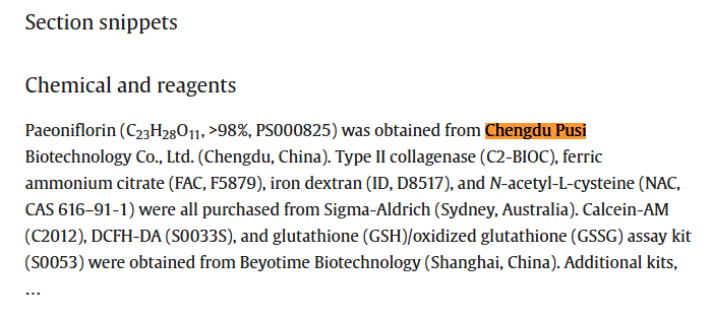
Wu B ,Luo Z ,Chen Z , et al.Paeoniflorin mitigates iron overload-induced osteoarthritis by suppressing chondrocyte ferroptosis via the p53/SLC7A11/GPX4 pathway.[J].International immunopharmacology,2025,162115111.
本文来自: 发布时间:2026-05-18
发表期刊:International Immunopharmacology
发表时间:2025
Abstract:
Background
Ferroptosis in chondrocytes is increasingly recognized as a key driver of osteoarthritis (OA) progression. Although paeoniflorin (PAE) has demonstrated potent anti-inflammatory and antioxidant properties in multiple disease models, its role in modulating OA through ferroptosis remains unclear.
Purpose
This study aimed to investigate the protective role of PAE against iron overload-induced OA (IOOA) via the p53/solute carrier family 7 member 11 (SLC7A11)/glutathione peroxidase 4 (GPX4) signaling pathway.
Methods
An iron overload model was established in chondrocytes using ferric ammonium citrate for in vitro experiments, while an in vivo IOOA model was induced in mice via destabilization of the medial meniscus combined with iron dextran injection. Subsequent evaluations included cell viability, cartilage matrix metabolism, iron accumulation, oxidative stress markers, and mitochondrial function. To investigate the underlying mechanism, Western blot, immunofluorescence (IF), and Nutlin-3 (a p53 activator) intervention were employed to assess the involvement of the p53/SLC7A11/GPX4 pathway. In vivo assessments included micro-computed tomography imaging, histological analysis, and IF.
Results
PAE significantly improved chondrocyte viability, restored matrix metabolism, reduced iron accumulation and oxidative stress, and protected mitochondrial function under iron overload conditions. Mechanistically, PAE downregulated p53 and upregulated SLC7A11 and GPX4 expression, thereby suppressing ferroptosis. Nutlin-3 partially reversed these protective effects. In vivo, PAE mitigated subchondral bone loss and cartilage destruction, reduced iron deposition, and restored GPX4 and type II collagen (COL2) expression while lowering matrix metalloproteinase 13 (MMP13) levels.
Conclusion
PAE alleviates iron overload-induced OA progression by inhibiting ferroptosis through regulation of the p53/SLC7A11/GPX4 pathway, offering new insights into ferroptosis-targeted OA therapy.
https://doi.org/10.1016/j.intimp.2025.115111

 关注官方微信